Ready to see more clearly?
If you have been diagnosed with keratoconus or suspect you may be affected, we are here to help. Book a specialist consultation and take the first step towards better vision.
What are the symptoms of Keratoconus?
Distorted vision
Straight lines may appear wavy or bent, and you may experience ghosting or multiple images. Difficulty getting glasses that work can also be a sign.
Sensitivity to light
Glare and haloes around lights are common, particularly noticeable when driving at night. This can often be described as "Double vision"
Changes in prescription
Rapid shifts in spectacle prescription, particularly increasing astigmatism, can be an early warning sign. Often standard contcat lenses do not work.
Allergies and eye rubbing
Soft contact lenses and spectacles often fail to adequately correct the irregular astigmatism keratoconus causes
What about other options?

Specialist Contact lenses
In our opinion mini-scleral lenses are the best lenses in the majority of cases, however, other lens forma are available should they be required. They include: Full scleral lenses — larger diameter than mini sclerals, used in more complex or advanced cases. Rose K / corneal RGP lenses — custom-designed rigid gas-permeable lenses that sit directly on the cornea. Effective but require an adaptation period. Hybrid lenses — a rigid centre with a soft skirt, offering some of the clarity of an RGP with improved comfort. Soft toric lenses — only suitable in very early or mild keratoconus where irregularity is minimal.

Collagen cross-linking (CXL)
Corneal collagen cross-linking is currently the only treatment that can halt the progression of keratoconus rather than simply correct the visual effects. It does not reverse existing damage, but it stabilises the cornea and prevents further deterioration. The procedure involves applying riboflavin (vitamin B2) eye drops to the corneal surface and then exposing the cornea to controlled ultraviolet-A light. This stimulates the formation of new bonds between collagen fibres, strengthening and stiffening the cornea. Cross-linking is typically recommended when keratoconus is shown to be progressing — usually identified through serial corneal topography measurements. It is performed as a day procedure by an ophthalmologist, most commonly under the NHS or privately at specialist corneal clinics.

Intrastromal Corneal Rings
Intrastromal corneal ring segments are small, crescent-shaped implants inserted into the peripheral cornea to reshape it and reduce the cone. They work by flattening the central cornea, which can improve both unaided and contact-lens-corrected vision. ICRS are a useful option for patients who are struggling to tolerate contact lenses or who wish to improve their uncorrected vision. They do not halt progression on their own and are often combined with cross-linking. The procedure is reversible — the segments can be removed or replaced if needed.

Phakic Intraocular Lenses (pIOL)
In some patients with keratoconus, particularly where the cornea is stable and the primary issue is a high refractive error (typically high myopia), a phakic intraocular lens may be implanted in front of the natural crystalline lens. This is a more complex surgical intervention and is not suitable for everyone, but can provide significant visual improvement in carefully selected cases.

Deep Anterior Lamellar Keratoplasty (DALK)
DALK is a form of partial corneal transplant in which the diseased anterior (front) layers of the cornea are removed and replaced with donor tissue, while the patient's own healthy inner layer (the endothelium) is preserved. Because the endothelium is retained, the risk of rejection is significantly lower than with a full-thickness transplant. DALK is generally considered when keratoconus has progressed to a stage where contact lenses can no longer provide functional vision, or where significant corneal scarring has developed.

Penetrating Keratoplasty (PK)
In the most advanced cases of keratoconus, particularly where central scarring is present and other surgical options are not viable, a full-thickness corneal transplant may be required. Penetrating keratoplasty involves replacing the entire central cornea with donor tissue. Graft survival rates are high, but recovery is lengthy — visual rehabilitation can take one to two years — and there is a lifelong risk of rejection. Specialist contact lenses, including scleral lenses, are often still required after transplant to achieve optimal vision.
TRY OUR VISION SIMULATOR
If you know your Kmax value, enter it below. If not, simply adjust the sliders until the simulated corneal topography closely resembles your own.
The Ray Trace Visualiser provides an approximation of how vision may be affected by keratoconus, helping to demonstrate the impact that corneal irregularity can have on image quality.
This can be a valuable way to help family, friends, or careers better understand what living with keratoconus is like. Because the condition affects vision in a complex and highly individual way, it can often be difficult to describe with words alone.
UNDERSTANDING ECTASIA OF THE CORNEA AND KERATOCONUS
What is Keratoconus?
Ectasia, also known as ectasis, is a term derived from the Greek word meaning "dilation" or "distention" of a tubular structure. In the context of the cornea, which is composed of a highly organised arrangement of collagen tubular fibers, ectasia refers to a group of conditions where this structural integrity is compromised. Such disruptions can lead to significant visual impairments. As the cornea accounts for approximately 90% of the eye's focusing power, any alteration to its structure can have a profound impact on the overall focusing ability of the visual system.
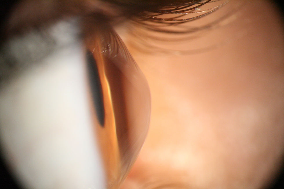
Keratoconus is the most common form of ectasia and is a progressive eye condition affecting the cornea — the clear, dome-shaped surface at the front of the eye. In a healthy eye the cornea is roughly spherical, but in keratoconus it gradually thins and bulges outward, forming an irregular cone shape. This distorts the way light enters the eye, causing increasingly blurred and distorted vision that spectacles alone can rarely fully correct.
It most commonly begins in the teenage years or early twenties and can progress into a person's thirties and forties. In more advanced cases, the corneal thinning can lead to scarring and, in severe instances, may require surgical intervention such as a corneal transplant. Early detection and careful management are therefore essential to preserving sight and quality of life.

Typical age of onset is between 10 and 25, and can usually to the age of 30. Risks include a family history, eye rubbing, and atopic (allergic) conditions like asthma, eczema, or hay fever.
Keratoconus

Pellucid Marginal Degeneration (PMD)
Usually presents in mid life between the ages of 20 and 40 but crucially can continue progress throughout life. Risks may include genetic predisposition, chronic eye rubbing and certain systemic conditions like connective tissue disorders.

Often starts asymptomatically in adulthood, usually between the ages of 20 and 40, and primarily affects men more than women. Risks are not well-known but may include a genetic predisposition and chronic inflammation.
Terriens

Typically present from birth (congenital) but can also develop later in life. Risk factors include genetic predisposition, connective tissue disorders such as Ehlers-Danlos syndrome or Marfan syndrome, and trauma to the eye.
Keratoglobus
THE SPECIALIST SOLUTION
What are mini scleral contact lenses?

Mini scleral lenses are a type of rigid gas-permeable (RGP) contact lens that are larger in diameter than a conventional corneal RGP lens but smaller than a full scleral lens — typically ranging from around 15 mm to 18 mm across. This intermediate size makes them an excellent middle ground: large enough to vault completely over an irregular cornea and rest on the white of the eye (the sclera), yet small and light enough to be comfortable for all-day wear.
Because the lens arches over the cornea without touching it, a reservoir of sterile saline fills the space between the back surface of the lens and the front surface of the eye. This fluid layer effectively replaces the irregular corneal surface with a smooth, regular one — giving light a consistent, unobstructed path into the eye and dramatically improving visual acuity.
Who can benefit?
Mini scleral lenses are particularly well suited to people with keratoconus who can no longer achieve adequate vision correction with spectacles or soft contact lenses, those who have found standard RGP lenses too uncomfortable, patients with other forms of corneal irregularity such as pellucid marginal degeneration or post-surgical corneas, and people who experience significant dry eye alongside corneal irregularity.
Key Benefits
-
Significantly improved clarity compared to spectacles or soft lenses
-
Complete corneal vaulting means no lens-on-cornea contact, reducing irritation
-
The saline reservoir provides continuous hydration, ideal for dry eye sufferers
-
Stable on the eye — less movement than standard RGP lenses
-
Suitable across a wide range of keratoconus severity
-
Custom designed and manufactured for your individual eye shape
-
Gas-permeable material ensures healthy oxygen supply to the cornea

YOUR JOURNEY
What to expect at your appointment
1. Initial consultation and corneal assessment
We begin with a thorough assessment of your eyes, including corneal topography — a detailed mapping of the shape of your cornea. This helps us understand the precise nature and severity of your keratoconus and determine whether mini scleral lenses are the most appropriate option for you.
2. Trial lens selection and fitting
Using your topography data, we select a trial lens that closely matches your eye's requirements. We apply the lens, fill the reservoir with saline, and carefully assess the fit using a slit lamp — checking the vault over the cornea, the landing zone on the sclera, and the overall centration and movement of the lens.
3. Over-refraction and vision assessment
Once the fit is confirmed, we check your vision through the lens and perform an over-refraction to determine the final prescription required. We want to ensure you achieve the best possible visual acuity before ordering your custom lenses.
4. Custom lens manufacture
Your lenses are manufactured to your individual specifications — incorporating your unique corneal shape, required prescription, and any specific design modifications identified during the fitting process. This typically takes one to two weeks.
5. Dispensing and insertion training
When your lenses arrive, we will show you how to insert and remove them correctly using the specialist plunger technique — slightly different from standard lens handling. We will not let you leave until you are confident doing this yourself.
6. Ongoing aftercare
Regular aftercare appointments are an important part of your care. We monitor the fit as your cornea changes over time, check the health of your eye, and make any adjustments needed to keep your vision and comfort at their best.
Frequently asked questions
What causes Keratoconus?
Keratoconus is a complex condition and one that researchers continue to study closely. While a definitive single cause has not been identified, several important risk factors and associations are now well established.
There is a modest genetic component, keratoconus can run in families, though the link is not strong enough to predict who will develop the condition. More consistently, keratoconus is associated with chronic ocular inflammation. Patients frequently have a history of allergies, hay fever, or systemic inflammatory conditions, and this pattern strongly suggests that ongoing inflammation plays a meaningful role in corneal weakening and disease progression.
One of the most clinically significant observations in keratoconus management is the strong association between eye rubbing and disease progression. Patients with keratoconus tend to rub their eyes frequently, though whether this is a cause or a consequence of the condition remains under investigation.
What is clear is this: stopping eye rubbing is one of the single most important steps a keratoconus patient can take to protect their corneas. Repeated mechanical pressure from rubbing accelerates corneal thinning and can worsen the cone shape over time.
Managing the underlying triggers, such as allergic conjunctivitis or hay fever, is therefore a key part of keratoconus care. This may involve antihistamine eye drops, mast cell stabilisers, or preservative-free ocular lubricants to reduce itching and the urge to rub. All of which we can advise you about at a consultation appointment.
The role of eye rubbing in Keratoconus.